fossil equine lower teeth
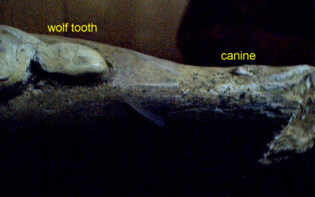
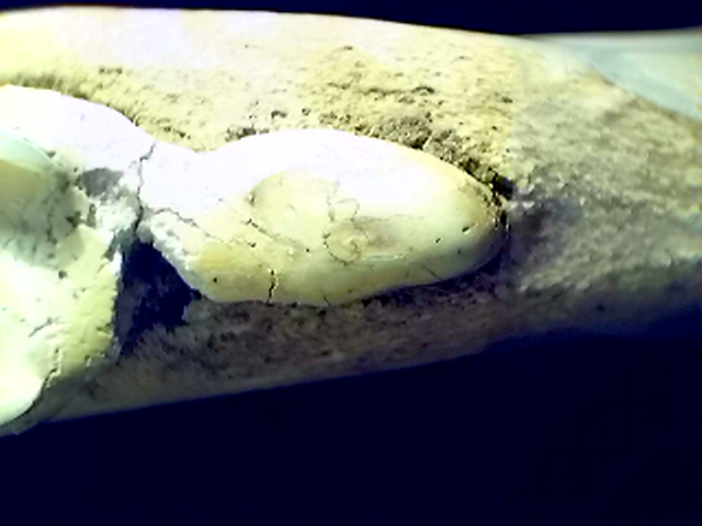
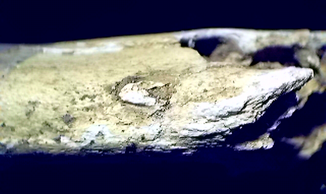
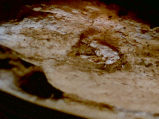
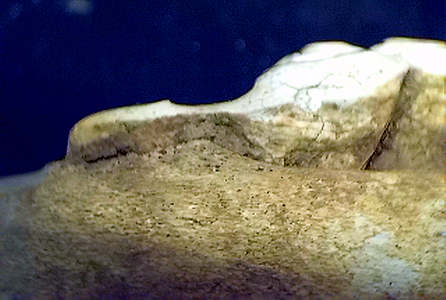
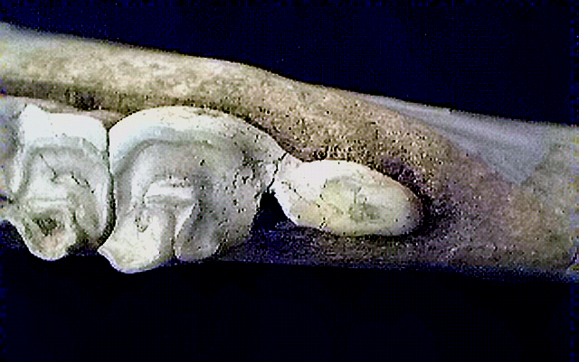
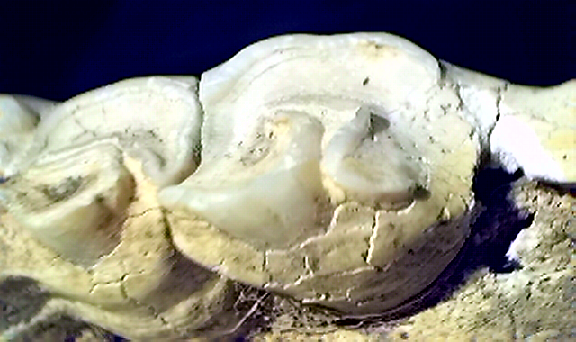
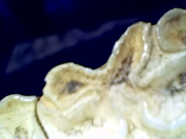
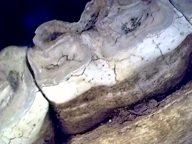
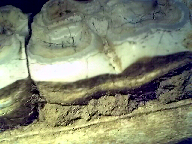
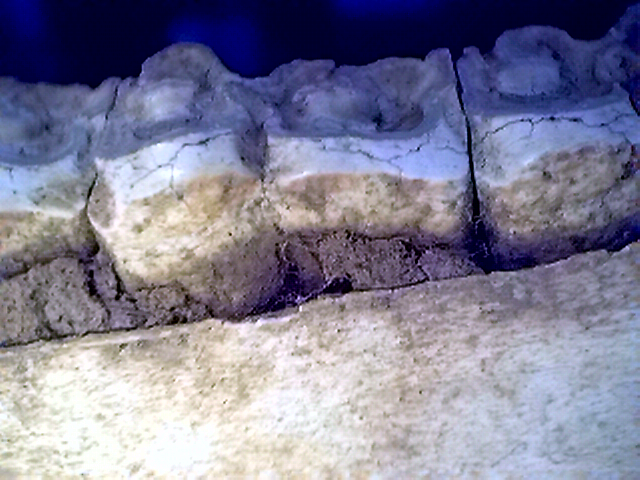
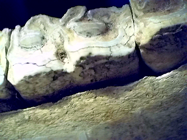
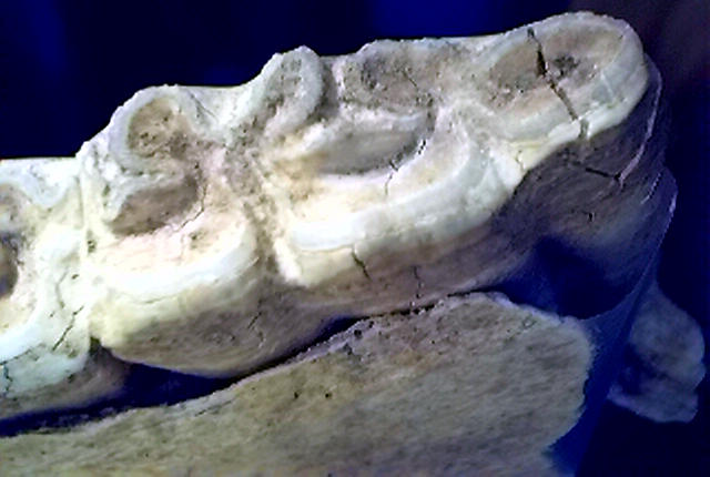
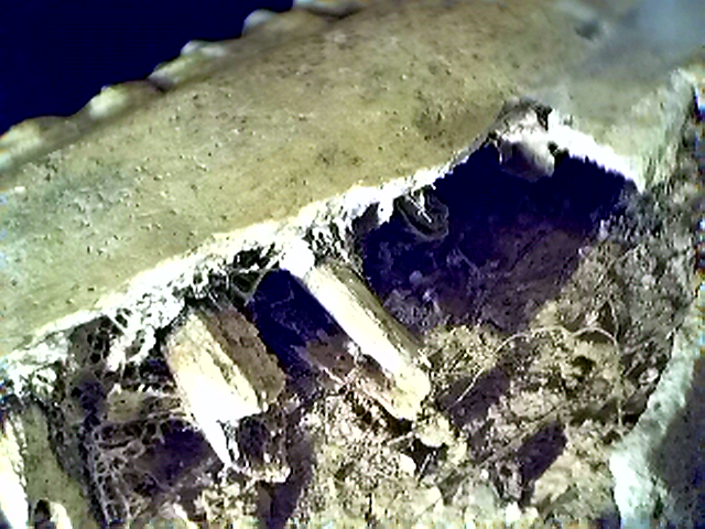
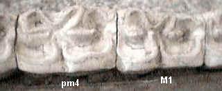
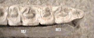
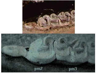
In 1996, I placed a number of teeth in my Hallquist collection.

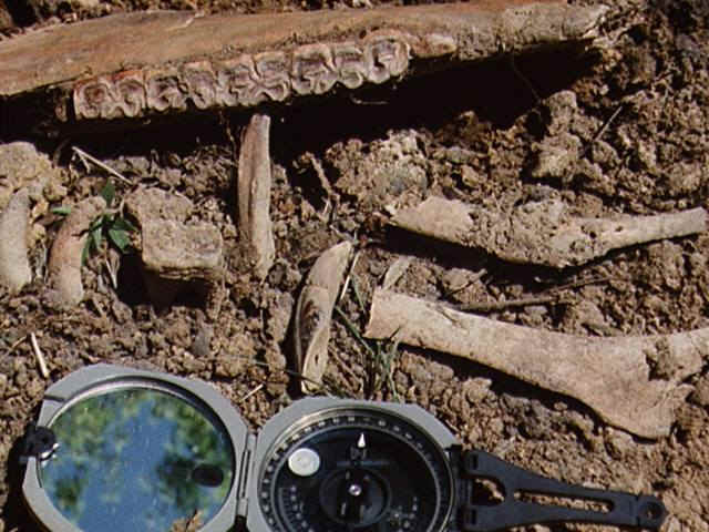
I added a tooth found near the row of five cheek teeth shown above to complete the row. Information on the Anatomy page of Equine Dentistry was used site to label the set as premolars pm2, pm3, pm4 and molars M1, M2, M3. I also learned from that site that the teeth are from a left side lower cheekbone.
Except for the image taken at the Hallquist discovery location, I used an Intel Create and Share PCI camera. I believe much better images would have been obtained if I had used my Olympus OM-1 35 mm film camera and a macro bellows arrangement.

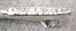
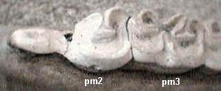
If you feel you are developing an interest in geology, please be prepared to experiment with image enhancement and comparison.

It would be helpful for you to own graphic software that will allow you to rotate, copy, cut and paste images. Be able to label images in the manner shown above for premolars and molars.

Concentrate on doing some detailed things well. Identify colleagues who will exchange and discuss features of images with you.

I started to examine the lower jaw specimen on April 19, 2009.

The lower jaw specimen exhibits the teeth and their roots. The root area is very complex. For example, there are fine fibers which would require many hours to document. There are clumps of sand adhering to the bone. I took one ignominious clump and crumbled it between two fingers. I deposited the resulting loose sand grains onto the QX3+ microscope stage. An image at 60X is shown. The material is loess: it was formed by wind blowing around the surface of a receding glacier.

our address: glennbowie@live.com.